Documented Scientific Record · 1888 – 1971

The Legacy of
Royal Raymond Rife

An American optical engineer and bacteriologist whose published research, institutional recognition, and instrument designs influenced microscopy for decades. This is the documented record — drawn only from verifiable primary sources.

A Documented Life

The Record in Chronological Order

Every entry on this timeline is drawn from published journal papers, official institutional reports, or verifiable historical documentation.

1913 – 1920

Optical Apprenticeship at Carl Zeiss

Rife trained in advanced microscopy and precision optics during his years at the Carl Zeiss optical works — then the most prestigious optical-instrument manufacturer in the world. He returned to San Diego with deep expertise in quartz-optic fabrication and polarized-light techniques.

1920s

Laboratory of Research Bacteriology, San Diego

Rife established his own research laboratory in San Diego and began his work on high-resolution optical microscopy — ultimately producing five generations of increasingly sophisticated instruments, each exceeding the optical limits thought possible at the time.

1931

First Peer-Reviewed Publication

Working with Professor Arthur Isaac Kendall of the Laboratory of Research Bacteriology at Northwestern University Medical School, Dr. Rife co-authored formal peer-reviewed research on filterable forms of Bacillus Typhosus. The paper was presented at the Bacteriological Section of the Los Angeles Clinical and Pathological Society on November 20, 1931.

Kendall, A.I. & Rife, R.R. · "Observations on Bacillus Typhosus in Its Filterable State" · California and Western Medicine · Vol XXXV · December 1931 · pp 409–411
1932

Published in Science (AAAS)

Independent confirmation of Rife's microscope capabilities appeared in Science, the flagship journal of the American Association for the Advancement of Science. Dr. E. C. Rosenow of the Mayo Clinic's Division of Experimental Bacteriology published observations using the Rife microscope.

Rosenow, E.C. · "Observations with the Rife microscope of filter-passing forms of microorganisms" · Science · Vol 76 · August 26, 1932 · pp 192–193
1933

The Universal Microscope Completed

The fifth and most advanced of Rife's microscopes — the Universal — was completed. Its achieved magnification (up to 17,000×) and resolving power far exceeded the theoretical limit for any contemporary light-based instrument, accomplished through a combination of quartz optics, polarized-light illumination, and Rife's distinctive sub-stage light source.

1944

Smithsonian Institution Recognition

A detailed technical article documenting Rife's microscope designs was published in the Annual Report of the Board of Regents of the Smithsonian Institution — one of the most respected institutional imprimaturs available to a scientific instrument of the era.

Seidel, R.E., M.D. & Winter, M. Elizabeth · "The New Microscopes" · Smithsonian Institution Annual Report · 1944 · pp 193–219
1944

Journal of the Franklin Institute

The same Seidel & Winter paper was published simultaneously in the Journal of the Franklin Institute — America's oldest continuously-operating scientific institute, and a body whose peer review was equivalent to that of any major engineering journal of the period.

Seidel & Winter · "The New Microscopes" · Journal of the Franklin Institute · Vol 237 · February 1944 · pp 103–130
1970s

Independent Verification by Modern Pathology

Decades later, Dr. John Hubbard — Associate Professor of Pathology at the State University of New York, Buffalo — formally reviewed the photographs Rife had published in 1944. Hubbard confirmed that the images demonstrated resolution approximately 20 angstroms, a capability modern light microscopy could not approach; the resolution was later corroborated by electron microscopy.

2013

Modern Peer-Reviewed Validation of Related Mechanisms

Research published in the Chinese Journal of Cancer demonstrated that specific low-intensity amplitude-modulated radiofrequency electromagnetic fields produce tissue- and tumor-specific effects in vitro — confirming, in modern peer-reviewed form, the underlying mechanism that Rife's era called "frequency specificity."

Zimmerman, Jimenez, Pennison, Brezovich, Morgan, Mudry, Costa, Barbault, Pasche · Chinese Journal of Cancer · 2013
Optical Engineering

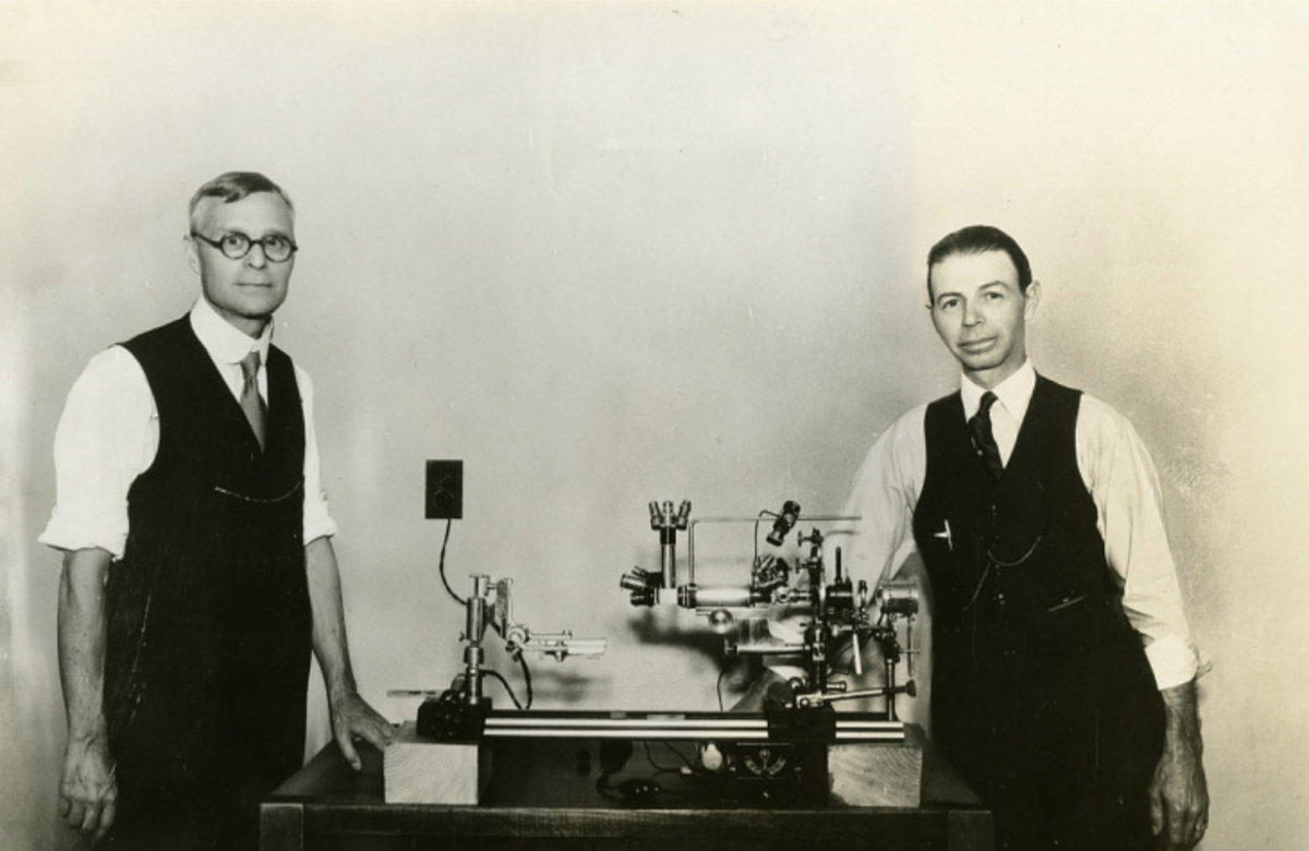
What Royal Rife Actually Invented

Setting aside every claim anyone else ever attached to his name, these are the instrumentation innovations that are Rife's alone — documented in patents, published papers, and surviving apparatus.
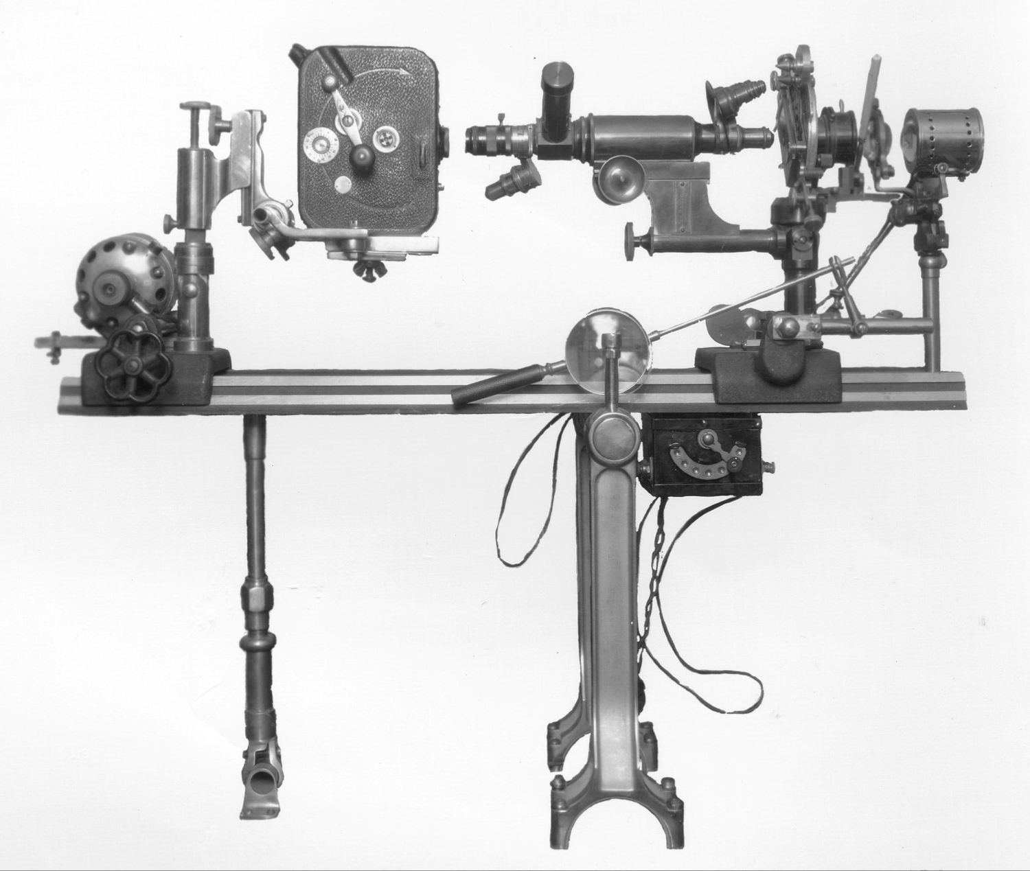
Dr. Rife's No. 1 Microscope — horizontal-stage architecture, brass and quartz construction, showing the integrated illumination, viewing column, and precision adjustment mechanisms
Microscope No. 1 — Horizontal-Stage Architecture Surviving instrument · circa late 1920s
Sub-Stage Illumination

The Rife Lamp

Rife designed a sub-stage illumination system that delivered extraordinarily intense, chromatically-controlled light to the specimen plane. The design was adopted by other microscope manufacturers of the era, including Otto Himmler of Berlin, who integrated Rife's lamp into his own precision instruments.

Quartz Optics

All-Quartz Optical Path

To transmit the full ultraviolet spectrum without absorption loss, Rife constructed microscope optical paths entirely from quartz — including the lenses, condenser, and illumination components. This was an engineering departure from the conventional glass optics used throughout the industry at the time.

Polarized Light

Double-Wedge Quartz Prism System

A double-wedge quartz prism mounted between the illuminating unit and the condenser, with vernier rotation through a full 360 degrees — allowing the observer to dial in any required angle of polarized light. This let Rife isolate optical properties of specimens that conventional microscopy could not resolve.

Resolution Beyond Diffraction

Overcoming the Fraunhofer Limit

Through the specific combination of quartz optics, polarized-light illumination, and his sub-stage lamp design, Rife's Universal Microscope achieved useful resolution down to approximately 20 angstroms — an accomplishment that would not be matched by any other light-based instrument in his lifetime, and was later confirmed by modern electron microscopy comparison.

Specimen Preparation

Frequency-Specific Response Methodology

Rife developed laboratory methods for identifying what he termed the "Mortal Oscillatory Rate" — a tissue- or pathogen-specific resonant frequency. Modern oncology research published in the Chinese Journal of Cancer (Zimmerman et al., 2013) has independently demonstrated that tumor-specific amplitude-modulated frequencies produce tissue-specific cellular effects — validating the underlying mechanism.

Horizontal Design

Horizontal-Stage Microscope Architecture

Unlike the vertical-column microscopes of his era, Rife's designs used a horizontal optical bench architecture allowing precise alignment of all components along a common axis. This architecture — unusual at the time — is echoed today in high-resolution imaging instruments requiring interferometric stability.

In The Words Of The Published Record

Direct From Peer-Reviewed Sources

Not paraphrased. Not interpreted. Directly from the journals and institutional reports of Dr. Rife's era.

Through the simultaneous availability of the Rife microscope, an instrument combining very high magnification with coördinated resolving power, and a simple procedure for inducing the filterable state in bacteria at will, the possibility of actually demonstrating organisms in this hitherto illusive condition very obviously presented itself.
California and Western Medicine · December 1931 Kendall, A.I. (Northwestern University) & Rife, R.R. — "Observations on Bacillus Typhosus in Its Filterable State"
The instrument is a universal microscope of high resolving power. Present-day microscopy is thus greatly extended, and it becomes possible for the first time in history to demonstrate visually the existence of the filtrable viruses.
Smithsonian Institution Annual Report · 1944 Seidel, R.E., M.D. & Winter, M. Elizabeth — "The New Microscopes"
The photographs that were published in the 1944 Smithsonian Report are indisputably beyond the capability of any resolution that was available at that time, and they have been confirmed by electron microscope since then. This has resolution down in the neighborhood of about 20 angstroms, at least, and nobody had ever been able to do that.
John Hubbard, M.D. · Assoc. Prof. of Pathology · SUNY Buffalo Modern verification of Rife's 1944 published photographs
Tumor-specific modulation frequencies regulate the expression of genes involved in migration and invasion and disrupt the mitotic spindle — demonstrating the ability of tumor-specific frequencies to block the growth of tumor cells in a tissue- and tumor-specific fashion.
Chinese Journal of Cancer · 2013 · Peer-reviewed Zimmerman et al. — Modern validation of frequency-specific cellular response
Collaborators & Peer Recognition

The Scientists Who Worked With Him

Dr. Rife was not a lone operator. His research was independently reviewed, replicated, and extended by named academic pathologists, bacteriologists, and physicians at respected institutions of his era.

Dr. Royal Raymond Rife (right) and Professor Arthur Isaac Kendall of Northwestern University Medical School (left), with one of Dr. Rife's horizontal microscopes
Dr. Royal Raymond Rife & Prof. Arthur Isaac Kendall — with the No. 1 Microscope From the Smithsonian Institution Collection · early 1930s
Northwestern University · Bacteriology

Professor Arthur Isaac Kendall, Ph.D.

Director of Research Bacteriology at Northwestern University Medical School. Co-authored the 1931 paper with Rife in California and Western Medicine. Kendall's independent use of the Rife microscope to confirm filter-passing bacterial forms was the earliest rigorous academic endorsement of Rife's optical work.

Mayo Clinic · Experimental Bacteriology

Edward C. Rosenow, M.D.

Of the Mayo Clinic's Division of Experimental Bacteriology — one of the most respected clinical research institutions in the world. Published independent confirmation of the Rife microscope's capabilities in Science (the AAAS journal) on August 26, 1932.

University of Southern California

Dr. Milbank Johnson, M.D.

Professor Emeritus of Physiological Therapeutics at USC College of Medicine, chairman of the AMA Southern California Division. Convened the USC Special Medical Research Committee to investigate Rife's work from 1931 onward, drawing physicians and pathologists from across the Southern California medical community.

USC · Pathology

Alvin G. Foord, M.D. & Others

Professor of Pathology at USC, and one of several named academic pathologists and physicians who participated in the USC Special Medical Research Committee's multi-year investigation of Rife's instruments and methods.

Franklin Institute · Engineering

R. E. Seidel, M.D. & M. Elizabeth Winter

Authored the comprehensive 1944 technical review of Rife's microscopes — first published in the Journal of the Franklin Institute (the United States' oldest continuously-operating scientific institute), then reprinted in the Annual Report of the Board of Regents of the Smithsonian Institution the same year.

SUNY Buffalo · Pathology

John Hubbard, M.D.

Associate Professor of Pathology at the State University of New York, Buffalo. In the 1970s, Hubbard formally reviewed the original photomicrographs published by Rife in the 1944 Smithsonian Report and confirmed resolution "down in the neighborhood of about 20 angstroms" — a capability that has since been verified by modern electron microscopy.

What Survives

The Innovations Still In Use

Many of Dr. Rife's documented contributions continue — sometimes under different names, sometimes incorporated into other instruments, but always traceable back to his published work.

From The Founder
My great-uncle was a real scientist. His instruments are in museum collections. His papers are in indexed medical journals. His work was confirmed by the Smithsonian. The abuse of his name by people he never met, decades after he died, does not erase what he actually did. Rife Systems exists to build in continuation of the documented record — and to give serious people a place to encounter the real man.
— Matthew Rife, Founder
Browse the Research Archive →